Coherent X-ray Scattering
|
Coherent X-ray Diffraction Imaging |
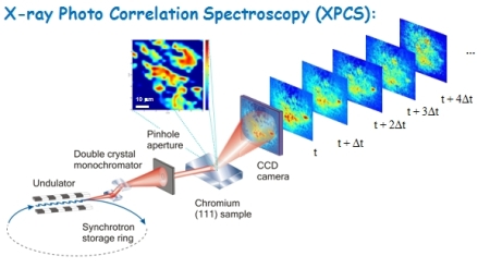
X-ray Photon Correlation Spectroscopy:
|
Coherent waves scattered from various objects are known to cause interference patterns, with one of the most famous examples - Young's double-slit interference experiment.
In case of double-slit experiment the interference pattern is periodic, with distance between two fringers related to the distance between slits and the width of the envelope defined by the size of the slit.
|
|
|
|
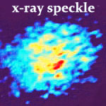
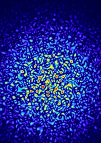
When one extends the same argument to a more complex configuration of random scatterers, the far-field diffraction pattern is a result of interference between various objects, a generally random "speckle" pattern, with size of the speckle related to the size of the coherent beam and width of the envelope defined by average size of the object, or correlation length in the scattering media.
Mathematically speaking, the speckle pattern is simply a Fourier Transform of the scattering objects (particles, domains, etc.) located within the illuminated volume. This provides a great sensitivity to even subtle motion of even a single scatterer, which modifies interference pattern.
The basic principle of X-ray Photon Correlation Spectroscopy (XPCS) is to study dynamics of scattering objects by means of observing time-dependent changes in the speckle pattern.
[top]
|
|
Laser speckle "in your kitchen":
|
Laser speckle images taken with a basic (cheap!) digital photo camera in transmission (left) and reflection (right) geometries. |
|
In addition to XPCS measurements which can only be performed at 3rd and 4th generation synchrotron sources, we are developing an in-house Dynamic Light Scattering setup, which uses visible light speckle produced by laser, and enables studies of dynamics of mesoscopic materials in the micron to centimeter lengthscale range.
One of the recent developments in Dynamic Light Scattering is the use of CCD (Charge Couple Device) detectors which enable study of slow and non-equilibrium processes. Use of two-detector detection scheme employed in a CCD also allows elimination of multiple scattering contributions which are one of the major obstacles in interpreting DLS results.
We are also exploring the use of Near-Field Speckle using both visible and x-ray scattering.
|
|
| [top] | |
|
Ptychographic reconstruction of famous Lenna image: artifacts and blurring disappear as the iterative algorithm progresses |
Coherent Diffractive Imaging, Phase Retrieval: A diffracted pattern is a Fourier transform of scattering density, characterized by phases and intensities. However, only intensities can be typically measured, while phase information is lost. In case the illuminated object is oversampled by at least a factor of 2, which means that the diffracted pattern is sampled at spatial frequencies at least twice that corresponding to the size of the sample, the phases can be recovered by algorithms. The oversampling criteria arises from information theory by Shannon (C. E. Shannon, C.E. Prec. Inst. Radio Engrs., N.Y. 37, 10 (1949)) and was first pointed out by Sayre ("Some Implications of a theorem due to Shannon", D. Sayre, Acta Cryst. 5 843 (1952)), and demonstrated for x-ray scattering by Miao et al. (J. Miao, P. Charalambous, J. Kirz, and D. Sayre, Nature (London) 400, 342 (1999)) |

|
One of the key advantage of this technique is that 3D microscopy of objects can be done without objective lenses, with resolution ultimately defined by the diffraction limit, rather than imperfection of the imaging optics.
Recent scientific highlights of lens-less imaging include the following references:
The lens less imaging algorithm can be applied to extended objects using ptychographic principle, which involves phase retrieval of multiple diffraction patterns obtained from overlapping regions on the sample.
Originally developed for electron microscopy (P. D. Nellist et al., Nature 374, 630 - 632 (2002), Hoppe, W. Acta crystallogr. A25, 495-501 (1969), H.M.L. Faulkner, J.M. Rodenburg, Phys. Rev. Lett., 93 023903 (2004); J.M. Rodenburg, H.M.L. Faulkner, Appl. Phys. Lett., 85 (4795), 2004), it has been recently demonstrated using visible (laser) light (
O. Bunk, M. Dierolf, S. Kynde, I. Johnson, O. Marti, and F. Pfeiffer,
Ultramicroscopy, 108, 481-487 (2007) and x-ray (J.M. Rodenburg et al.,
Phys. Rev. Lett. 98, 034801 (2007)).
Our group is currently developing application of ptychographic imaging in both x-ray and visible light scattering regimes.
Fresnel Coherent Diffractive Imaging The lens less imaging algorithm can be applied to extended objects using ptychographic principle, which involves phase retrieval of multiple diffraction patterns obtained from overlapping regions on the sample.
Another direction we are currently pursuing is Fresnel Coherent Diffractive Imaging technique, where speckle is recorded in near-field (Fresnel) regime, rather than far-field (Fraunhoffer).
|
|
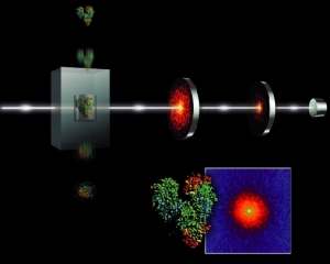
One of the intriguing future applications of Coherent X-ray Diffractive Imaging is the possibility to determine atomic structure of individual molecules - such as hard-to-crystallize biological proteins. Biological materials become easily damaged and destroyed by powerful x-ray beams, however, incredibly short femtosecond x-ray pulses produced by newly available X-ray Free Electron Lasers may enable scientists to image molecules and nanoparticles with angstrom-level resolution before they disintegrate.
For more on these directions see the following references: |
|
